USMLE Reviewer
(By Subscription)
Update October 31, 2019
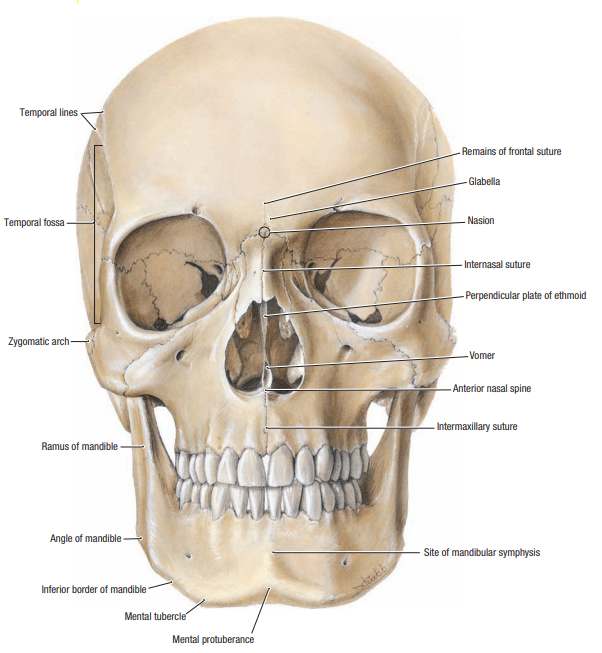
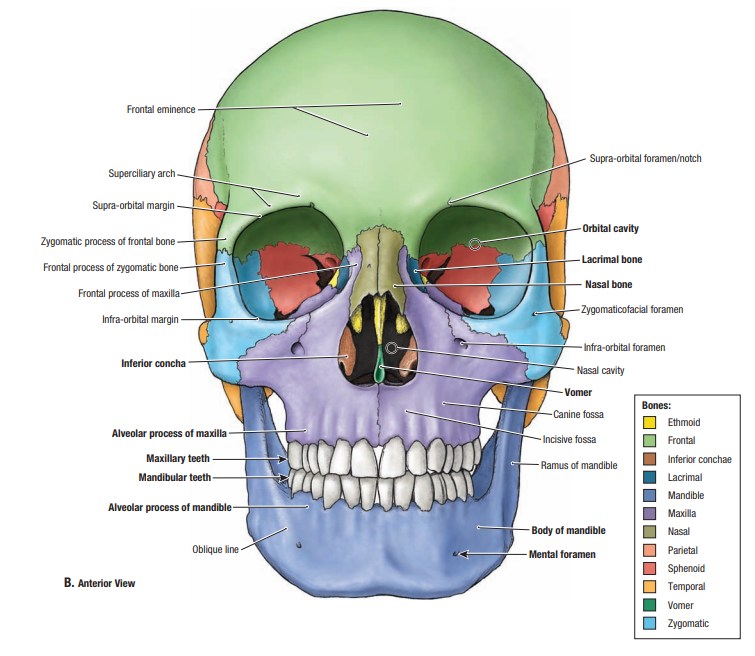
The skull consists of bones that protect the brain.
The cranial bones consist of spongy bone “sandwiched” between two layers of compact bone.
The outer and inner surfaces of the skull, are covered by periosteum, known respectively as the pericranium and the endocranium.
The periosteum is continuous at the sutures of the skull.
Hover on Label for more Information
Click on Label for More Information
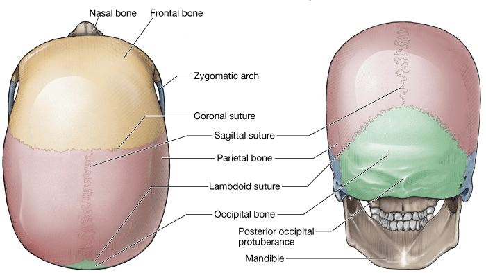
Sutures are immovable fibrous joints between the bones of the skull. The principal sutures are:
Clinical Correlation
The anterior division of the middle meningeal artery courses deep to the pterion on the inner surface of the skull. A blow to the pterion, therefore, could rupture the middle meningeal artery and result in an epidural (extradural) hematoma, which is a buildup of blood between the dura mater and the skull. On a radiograph, an epidural hematoma appears as a convex shape because sutures at the sites where the periosteal dura is more firmly attached to the skull stop the hematoma from expanding. As a result, epidural hematomas expand inward toward the brain instead of along the side of the skull, which occurs in a subdural hematoma. Unconsciousness and death occur rapidly because of the bleeding that dissects a wide space as it strips the dura from the inner surface of the skull, causing pressure on the brain.
(By Subscription)