Meiosis I

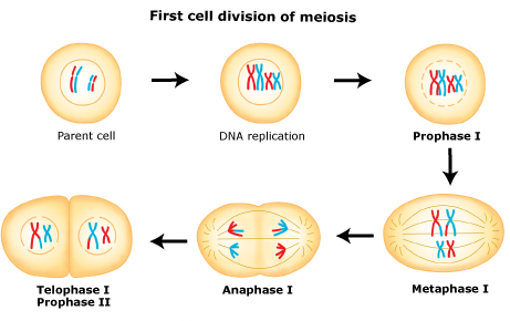
In meiosis I, homologous chromosomes pair with each other.

Oogonia undergo meiosis in their transformation to oocytes but arrest at prophase of meiosis I until the time of ovulation at puberty, age 9-10 when it resumes.

This stage of development is called the dictyate. In the dictyate (prolonged diplotene) stage actively repairs DNA damage.

Also at this stage the chromosomes can exchange genetic material in a process called chromosomal crossover. The homologous chromosomes are then pulled apart into two new separate daughter cells, each containing half the number of chromosomes as the parent cell. Thus meiosis I is referred to as a reductional division. At the end of meiosis I, sister chromatids remain attached and may differ from one another if crossing-over occurred.

A regular diploid human cell contains 46 chromosomes and is considered 2N because it contains 23 pairs of homologous chromosomes. However, after meiosis I, although the cell contains 46 chromatids, it is only considered as being N, with 23 chromosomes. This is because in Anaphase I the sister chromatids will remain together as the spindle fibers pull the pair toward the pole of the new cell.


Meiosis and Diversity in Gene Expression

Meiosis generates gamete genetic diversity in two ways:

(1) the independent orientation of homologous chromosome pairs along the metaphase plate during metaphase I and the subsequent separation of homologs during anaphase I allows a random and independent distribution of chromosomes to each daughter cell (and ultimately to gametes); and

(2) physical exchange of homologous chromosomal regions by homologous recombination during prophase I results in new combinations of DNA within chromosomes.

Chromosomal crossover (or crossing over) is the exchange of genetic material between homologous chromosomes that results in recombinant chromosomes. It is one of the final phases of genetic recombination, which occurs during prophase I of meiosis during a process called synapsis. Synapsis begins before the synaptonemal complex develops, and is not completed until near the end of prophase I. Crossover usually occurs when matching regions on matching chromosomes break and then reconnect to the other chromosome. This process begins in early stage of prophase I which is called leptotene.1

Crossing over was described, in theory, by Thomas Hunt Morgan. He relied on the discovery of the Belgian Professor Frans Alfons Janssens of the University of Leuven who described the phenomenon in 1909 and had called it "chiasmatypie". The term chiasma is linked if not identical to chromosomal crossover. Morgan immediately saw the great importance of Janssens' cytological interpretation of chiasmata to the experimental results of his research on the heredity of Drosophila. The physical basis of crossing over was first demonstrated by Harriet Creighton and Barbara McClintock in 1931.[2]

In meiosis II, the two cells produced during meiosis I divide again. During this division, sister chromatids detach from one another and are separated into four total daughter cells. The daughter cells are haploid and contain only one copy of each chromosome.


Reference

1. http://en.wikipedia.org/wiki/Chromosomal_crossover


At this stage, the primary oocyte is surrounded by a single layer of flattened ovarian follicular epithelial cells. These cells are also known as granulosa cells).


Resumption of Meiosis I at Puberty

At puberty, a small number of primary oocytes (20-50) mature each month and complete the first meiotic division to become secondary oocytes, under the influence of follicle stimulating hormone. (See the hypothalamus-pituitary-gonadal axis). The menstrual cycle begins.

Primary oocytes continue to develop in each menstrual cycle. Synapsis occurs and tetrads form, enabling chromosomal crossover to occur.

At the end of meiosis I, the primary oocyte has now developed into the secondary oocyte and the first polar body.

Meiosis II

Immediately after the completion of meiosis I, the haploid secondary oocyte initiates meiosis II. However, this process is also halted at the metaphase II stage until fertilization, if such should ever occur. If the egg is not fertilized, it is disintegrated and released (menstruation) and the secondary oocyte does not complete meiosis II (and doesn't become an ovum). When meiosis II has completed, an ootid and another polar body have now been created.


After a second mitotic division, ova are formed.

The oocytes synthesise a coat and cortical granules - this glycoprotein coat is called the 'zona pellucida'. They also accumulate ribosomes, yolk, glycogen, lipid and the mRNA that will be used later on after fertilisation to direct early development of the embryo.

++++++++++++++++++++

Once you reach puberty, the ovaries release a single egg each month (the ovaries typically alternate releasing an egg)—this is called ovulation. The hypothalamus sends a signal to the pituitary gland to release gonadotrophic substances (follicle stimulating hormone and luteinizing hormone). These hormones are essential to normal reproductive function—including regulation of the menstrual cycle.

Meiosis

The egg cell remains as a primary ovum until the time for its release from the ovary arrives. The egg then undergoes a  cell division. The nucleus splits so that half of its chromosomes go to one cell and half to another. One of these two new cells is usually larger than the other and is known as the  secondary ovum; the smaller cell is known as a  polar body. The secondary ovum grows in the ovary until it reaches maturation; it then breaks loose and is carried into the fallopian tubes. Once in the fallopian tubes, the secondary egg cell is suitable for fertilization by the male sperm cells. See also ovulation; ovum.

++++++++++++++


Estrogen (estradiol, specifically) is instrumental in breast development, fat distribution in the hips, legs, and breasts, and the development of reproductive organs.
 
To a lesser extent, the ovaries release the hormone relaxin prior to giving birth. Another minor hormone is inhibin, which is important for signaling to the pituitary to inhibit follicle-stimulating hormone secretion.
 
Progesterone and Estrogen Production and Function

Progesterone and estrogen are necessary to prepare the uterus for menstruation, and their release is triggered by the hypothalamus.


As the egg migrates down the fallopian tube, progesterone is released. It is secreted by a temporary gland formed within the ovary after ovulation called the corpus luteum. Progesterone prepares the body for pregnancy by causing the uterine lining to thicken. If a woman is not pregnant, the corpus luteum disappears.
 
If a woman is pregnant, the pregnancy will trigger high levels of estrogen and progesterone, which prevent further eggs from maturing. Progesterone is secreted to prevent uterine contractions that may disturb the growing embryo. The hormone also prepares the breasts for lactation.
 
Increased estrogen levels near the end of pregnancy alert the pituitary gland to release oxytocin, which causes uterine contractions. Before delivery, the ovaries release relaxin, which as the name suggests, loosens the pelvic ligaments in preparation for labor.
 
More hormones are released during pregnancy than at any other time of a woman’s life, but during menopause—which marks the end of fertility—estrogen levels fall fast.
+++++++++++++++++++++


The oocytes are surrounded by epithelial cells and form follicles. The ovary contains many primordial follicles, which are mostly found around the edges of the cortex. There are fewer follicles in different stages of development.


The ovary produces both oocytes and sex hormones.

A diagram of a sectioned ovary (a) shows the different stages of follicle maturation, ovulation, and corpus luteum formation and degeneration. All of the stages and structures shown in this diagram actually would appear at different times during the ovarian cycle and do not occur simultaneously. Follicles are arranged here for easy comparisons. The primordial follicles shown are greatly enlarged. The histologic sections identify primordial follicles (b), a primary follicle (c), a secondary follicle (d), and a large vesicular follicle (e). After ovulation, the portion of the follicle left behind forms the corpus luteum (f), which then degenerates into the corpus albicans (g). All H&E.

  • Structure
  • Function


  • The ovary’s cortex is covered by a cuboidal mesothelium, the surface epithelium (or germinal epithelium) that overlies a layer of connective tissue, the tunica albuginea.

  • Before puberty all follicles are primordial follicles, formed in the developing fetal gonad, with each having one primary oocyte arrested in meiotic prophase I and a surrounding layer of squamous follicular epithelial cells.

  • After puberty some primordial follicles develop each month as growing primary follicles, with an enlarging primary oocyte surrounded by larger epithelial cells now called granulosa cells.

  • During follicular growth the granulosa cells, surrounded by a basement membrane, become stratified and actively engage in fluid secretion and steroid hormone metabolism.

  • Between the oocyte and the granulosa cells a thin layer forms called the zona pellucida, which contains glycoproteins (ZO proteins) to which the sperm surface must bind to reach the oocyte at fertilization.

  • Antral or vesicular follicles are larger and have developed fluid-filled spaces among their granulosa cells, but the growing oocyte is still in prophase I.

  • While the primary follicle grows, mesenchymal cells immediately around it form the highly vascular layer, the theca interna, and a more fibrous theca externa, with smooth muscle cells.

  • Endocrine cells of the theca interna secrete both progesterone and estrogen precursors, which are converted by granulosa cells into estrogen.

  • Antral follicles continue developing as mature, graafian follicles, which have a large antrum filled with fluid, with the large primary oocyte enclosed by granulosa cells of the cumulus oophorus.

  • Each month only one graafian follicle becomes a dominant follicle and undergoes ovulation; most other developing follicles arrest and degenerate with apoptosis in a process called atresia.


Throughout the ovarian cycle, a selected follicle is stimulated to undergo growth and development, culminating in ovulation. The remnants of the follicle undergo reorganization into thecorpus luteum, a temporary endocrine organ that plays a central role in preparation and maintenance of the initial stages of pregnancy. Parallel changes occur in endometrial morphology and function throughout the ovarian cycle in preparation for implantation of a fertilized ovum. Ovarian and placental hormones maintain pregnancy and prepare the breast for lactation. This chapter describes the basic principles of the neuroendocrine regulation of this hypothalamic-pituitary-ovarian axis.The ovaries are both a gonad and an endocrine gland. The ovaries produce eggs (ovum or oocytes) and sex hormones (e.g., estrogen).

  • As Gonad
  • As Endocirine Gland

Control of menstrual cycle


Ovulatory menstrual cycles are regulated by interactions of the hypothalamic-pituitary axis, ovaries, and endometrium.

Hypothalamus Pituitary Ovary Endometrium
Anterior and medial hypothalamus; preoptic septal areas Luteinizing hormone-releasing hormone Luteinizing hormone and follicle-stimulating hormone Gonadotroph


Image not available.


Gonadotropin control of the ovarian and endometrial cycles.

The ovarian-endometrial cycle has been structured as a 28-day cycle. The follicular phase (days 1 to 14) is characterized by rising estrogen levels, endometrial thickening, and selection of the dominant “ovulatory” follicle. During the luteal phase (days 14 to 21), the corpus luteum (CL) produces estrogen and progesterone, which prepare the endometrium for implantation. If implantation occurs, the developing blastocyst begins to produce human chorionic gonadotropin (hCG) and rescues the corpus luteum, thus maintaining progesterone production. FSH = follicle-stimulating hormone; LH = luteinizing hormone.

The average cycle duration is approximately 28 days, with a range of 25 to 32 days. The hormonal sequence leading to ovulation directs this cycle. Concurrently, cyclical changes in endometrial histology are faithfully reproduced.

Rock and Bartlett (1937) first suggested that endometrial histological features were sufficiently characteristic to permit cycle “dating.” In this scheme, the follicular-proliferative phase and the postovulatory luteal-secretory phase are customarily divided into early and late stages. These changes are detailed in Chapter 15 of Williams Gynecology, 2nd edition (Halvorson, 2012).

Follicular or Preovulatory Ovarian Phase

The human ovary contains 2 million oocytes at birth, and approximately 400,000 follicles are present at puberty onset (Baker, 1963). The remaining follicles are depleted at a rate of approximately 1000 follicles per month until age 35, when this rate accelerates (Faddy, 1992). Only 400 follicles are normally released during female reproductive life. Therefore, more than 99.9 percent of follicles undergo atresia through a process of cell death termed apoptosis (Gougeon, 1996; Kaipia, 1997).

Follicular development consists of several stages, which include the gonadotropin-independent recruitment of primordial follicles from the resting pool and their growth to the antral stage. This appears to be controlled by locally produced growth factors. Two members of the transforming growth factor-β family—growth differentiation factor 9 (GDF9) and bone morphogenetic protein 15 (BMP-15)—regulate granulosa cell proliferation and differentiation as primary follicles grow (Trombly, 2009; Yan, 2001). They also stabilize and expand the cumulus oocyte complex in the oviduct (Hreinsson, 2002). These factors are produced by oocytes, suggesting that the early steps in follicular development are, in part, oocyte controlled. As antral follicles develop, surrounding stromal cells are recruited, by a yet-to-be-defined mechanism, to become thecal cells.

Although not required for early follicular maturation, follicle-stimulating hormone (FSH) is required for further development of large antral follicles (Hillier, 2001). During each ovarian cycle, a group of antral follicles, known as a cohort, begins a phase of semisynchronous growth based on their maturation state during the FSH rise in the late luteal phase of the previous cycle. This FSH rise leading to follicle development is called the selection window of the ovarian cycle (Macklon, 2001). Only the follicles progressing to this stage develop the capacity to produce estrogen.

During the follicular phase, estrogen levels rise in parallel to growth of a dominant follicle and to the increase in its number of granulosa cells (see Figure 1. These cells are the exclusive site of FSH receptor expression. The rise of circulating FSH levels during the late luteal phase of the previous cycle stimulates an increase in FSH receptors and subsequently, the ability of cytochrome P450 aromatase within granulosa cells to convert androstenedione into estradiol. The requirement for thecal cells, which respond to luteinizing hormone (LH), and granulosa cells, which respond to FSH, represents the two-gonadotropin, two-cell hypothesis for estrogen biosynthesis (Short, 1962). As shown in Figure 5-2, FSH induces aromatase and expansion of the antrum of growing follicles. The follicle within the cohort that is most responsive to FSH is likely to be the first to produce estradiol and initiate expression of LH receptors.


The two-cell, two-gonadotropin principle of ovarian steroid hormone production. During the follicular phase (left panel), luteinizing hormone (LH) controls theca cell production of androstenedione, which diffuses into the adjacent granulosa cells and acts as precursor for estradiol biosynthesis. The granulosa cell capacity to convert androstenedione to estradiol is controlled by follicle-stimulating hormone (FSH). After ovulation (right panel), the corpus luteum forms and both theca-lutein and granulosa-lutein cells respond to LH. The theca-lutein cells continue to produce androstenedione, whereas granulosa-lutein cells greatly increase their capacity to produce progesterone and to convert androstenedione to estradiol. LH and hCG bind to the same LH-hCG receptor. If pregnancy occurs (right panel), human chorionic gonadotropin (hCG) rescues the corpus luteum through their shared LH-hCG receptor. Low-density lipoprotein (LDL) is an important source of cholesterol for steroidogenesis. cAMP = cyclic adenosine monophosphate.

+Image not available.


The ovaries produce steroidal sex hormones that control organs of the reproductive system and influence other organs. Beginning at menarche, when the first menses occurs, the reproductive system undergoes monthly changes in structure and function that are controlled by neurohormonal mechanisms. Menopause is a variably timed period during which the cyclic changes become irregular and eventually disappear. In the postmenopausal period the reproductive organs slowly involute. Although the mammary glands do not belong to the genital system, they are included here because they undergo changes directly connected to the functional state of the reproductive organs.


See: 04b. Hypothalamic–Pituitary–Ovarian Axis


  • Ovarian-Endometrial Menstrual Cycle

Ovulatory menstrual cycles are regulated by interactions of the hypothalamic-pituitary axis, ovaries, and endometrium.

Hypothalamus Pituitary Ovary Endometrium
Anterior and medial hypothalamus; preoptic septal areas Luteinizing hormone-releasing hormone Luteinizing hormone and follicle-stimulating hormone Gonadotroph


Image not available.


Gonadotropin control of the ovarian and endometrial cycles.

The ovarian-endometrial cycle has been structured as a 28-day cycle. The follicular phase (days 1 to 14) is characterized by rising estrogen levels, endometrial thickening, and selection of the dominant “ovulatory” follicle. During the luteal phase (days 14 to 21), the corpus luteum (CL) produces estrogen and progesterone, which prepare the endometrium for implantation. If implantation occurs, the developing blastocyst begins to produce human chorionic gonadotropin (hCG) and rescues the corpus luteum, thus maintaining progesterone production. FSH = follicle-stimulating hormone; LH = luteinizing hormone.

The average cycle duration is approximately 28 days, with a range of 25 to 32 days. The hormonal sequence leading to ovulation directs this cycle. Concurrently, cyclical changes in endometrial histology are faithfully reproduced.

Rock and Bartlett (1937) first suggested that endometrial histological features were sufficiently characteristic to permit cycle “dating.” In this scheme, the follicular-proliferative phase and the postovulatory luteal-secretory phase are customarily divided into early and late stages. These changes are detailed in Chapter 15 of Williams Gynecology, 2nd edition (Halvorson, 2012).

Follicular or Preovulatory Ovarian Phase

The human ovary contains 2 million oocytes at birth, and approximately 400,000 follicles are present at puberty onset (Baker, 1963). The remaining follicles are depleted at a rate of approximately 1000 follicles per month until age 35, when this rate accelerates (Faddy, 1992). Only 400 follicles are normally released during female reproductive life. Therefore, more than 99.9 percent of follicles undergo atresia through a process of cell death termed apoptosis (Gougeon, 1996; Kaipia, 1997).

Follicular development consists of several stages, which include the gonadotropin-independent recruitment of primordial follicles from the resting pool and their growth to the antral stage. This appears to be controlled by locally produced growth factors. Two members of the transforming growth factor-β family—growth differentiation factor 9 (GDF9) and bone morphogenetic protein 15 (BMP-15)—regulate granulosa cell proliferation and differentiation as primary follicles grow (Trombly, 2009; Yan, 2001). They also stabilize and expand the cumulus oocyte complex in the oviduct (Hreinsson, 2002). These factors are produced by oocytes, suggesting that the early steps in follicular development are, in part, oocyte controlled. As antral follicles develop, surrounding stromal cells are recruited, by a yet-to-be-defined mechanism, to become thecal cells.

Although not required for early follicular maturation, follicle-stimulating hormone (FSH) is required for further development of large antral follicles (Hillier, 2001). During each ovarian cycle, a group of antral follicles, known as a cohort, begins a phase of semisynchronous growth based on their maturation state during the FSH rise in the late luteal phase of the previous cycle. This FSH rise leading to follicle development is called the selection window of the ovarian cycle (Macklon, 2001). Only the follicles progressing to this stage develop the capacity to produce estrogen.

During the follicular phase, estrogen levels rise in parallel to growth of a dominant follicle and to the increase in its number of granulosa cells (see Figure 1. These cells are the exclusive site of FSH receptor expression. The rise of circulating FSH levels during the late luteal phase of the previous cycle stimulates an increase in FSH receptors and subsequently, the ability of cytochrome P450 aromatase within granulosa cells to convert androstenedione into estradiol. The requirement for thecal cells, which respond to luteinizing hormone (LH), and granulosa cells, which respond to FSH, represents the two-gonadotropin, two-cell hypothesis for estrogen biosynthesis (Short, 1962). As shown in Figure 5-2, FSH induces aromatase and expansion of the antrum of growing follicles. The follicle within the cohort that is most responsive to FSH is likely to be the first to produce estradiol and initiate expression of LH receptors.


The two-cell, two-gonadotropin principle of ovarian steroid hormone production. During the follicular phase (left panel), luteinizing hormone (LH) controls theca cell production of androstenedione, which diffuses into the adjacent granulosa cells and acts as precursor for estradiol biosynthesis. The granulosa cell capacity to convert androstenedione to estradiol is controlled by follicle-stimulating hormone (FSH). After ovulation (right panel), the corpus luteum forms and both theca-lutein and granulosa-lutein cells respond to LH. The theca-lutein cells continue to produce androstenedione, whereas granulosa-lutein cells greatly increase their capacity to produce progesterone and to convert androstenedione to estradiol. LH and hCG bind to the same LH-hCG receptor. If pregnancy occurs (right panel), human chorionic gonadotropin (hCG) rescues the corpus luteum through their shared LH-hCG receptor. Low-density lipoprotein (LDL) is an important source of cholesterol for steroidogenesis. cAMP = cyclic adenosine monophosphate.

+Image not available.


The ovaries produce steroidal sex hormones that control organs of the reproductive system and influence other organs. Beginning at menarche, when the first menses occurs, the reproductive system undergoes monthly changes in structure and function that are controlled by neurohormonal mechanisms. Menopause is a variably timed period during which the cyclic changes become irregular and eventually disappear. In the postmenopausal period the reproductive organs slowly involute. Although the mammary glands do not belong to the genital system, they are included here because they undergo changes directly connected to the functional state of the reproductive organs.


Folliculogenesis[edit]

Main article: Folliculogenesis

Synchronously with ootidogenesis, the ovarian follicle surrounding the ootid has developed from a primordial follicle to a preovulatory one.

Maturation into ovum[edit]

Both polar bodies disintegrate at the end of Meiosis II, leaving only the ootid, which then eventually undergoes maturation into a mature ovum.

The function of forming polar bodies is to discard the extra haploid sets of chromosomes that have resulted as a consequence of meiosis.

In vitro maturation[edit]

Main article: In vitro maturation

In vitro maturation (IVM) is the technique of letting ovarian follicles mature in vitro. It can potentially be performed before an IVF. In such cases, ovarian hyperstimulation isn't essential. Rather, oocytes can mature outside the body prior to IVF. Hence, no (or at least a lower dose of) gonadotropins have to be injected in the body.[20] Immature eggs have been grown until maturation in vitro at a 10% survival rate, but the technique is not yet clinically available.[21] With this technique, cryopreserved ovarian tissue could possibly be used to make oocytes that can directly undergo in vitro fertilization.[21]

++++++++++++++++++++++++++++++++++++++++


[By the time that the female is born, all of the egg cells that the ovaries will release during the active reproductive years of the female are already present in the ovaries. These cells number around 400,000.]


USMLE Reviewer (By Subscription)

Female Reproductive System

Endocrine System

Digital World Medical School
© 2020